


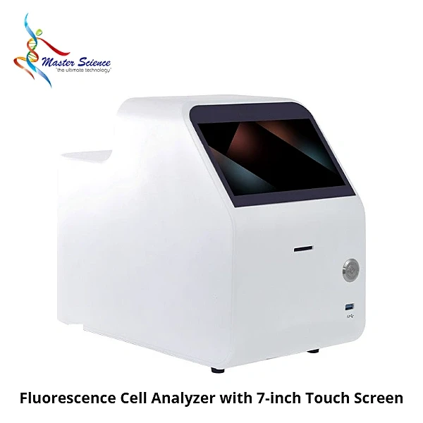
Fluorescence Cell Analyzer, Dual-Channel AO/PI Cell Counter, 5-200 μm, 6-Sample Autofocus
Inhouse product

The Master Science Fluorescence Cell Analyzer combines bright field imaging with two fluorescence excitation wavelengths and two detection filters for routine analysis of cell density, viability and transfection efficiency. It measures cells from 5 to 200 μm at concentrations of 1 x 10^4 to 3 x 10^7 cells/mL across a 0-100% viability range.
Autofocus, a 6.3 MP industrial CMOS camera and a six-chamber counting slide let it capture five fields of view per sample and process up to six samples per run. Pre-configured apps on the 7-inch multi-touch display guide each test.
Technical Specifications
Specifications of the Fluorescence Cell Analyzer:
- Brand: Master Science
- Model Number: MSFACC-TugerSWISS
- SKU: MSFACC-TugerSWISS
- Concentration Range: 1 x 10^4 to 3 x 10^7 cells/mL
- Cell Diameter Range: 5-200 μm
- Viability Range: 0-100%
- Sample Volume: 10 μL (GPC100, GR100) / 20 μL (GPC200, GR200)
- Analysis Time, Single Sample, Single View: Bright field < 1 s; fluorescence < 12 s (AO/PI)
- Analysis Time, Single Sample, Five Views: Bright field < 8 s; fluorescence < 54 s (AO/PI)
- Analysis Time, Six Samples, Five Views: Bright field < 55 s; fluorescence < 334 s (AO/PI)
- Excitation Wavelengths: 475 nm, 545 nm
- Emission Wavelengths: 530 nm, 605 nm
- Fluorescence Light Source: Blue LED / green LED
- Bright Field Light Source: White LED
- Output Formats: JPG, PDF, CSV; FCS optional
- Screen: 7-inch multi-touch colour display
- Camera: Industrial-grade colour CMOS, 6.3 MP
- Storage: 128 GB standard (other capacities optional)
- Focus: Autofocus
- Interfaces: 2 x USB, 1 x HDMI, 1 x RJ45
- Consumable: Six-chamber counting slide
- Dimensions (W x D x H): 190 x 276 x 367 mm
- Weight: 4.4 kg (without power adapter)
Key Benefits
- Two fluorescence channels: Supports AO, PI, AO/PI, GFP and RFP assays.
- Higher throughput: Six samples per slide with five fields of view each.
- Clear images automatically: Autofocus with a 6.3 MP colour CMOS camera.
- Data integrity tools: Multi-level user permissions, encrypted export, electronic signatures and audit trails.
For bright field and fluorescence imaging of cultures, see our Inverted Fluorescence Microscope, 3-Channel LED next to the Fluorescence Cell Analyzer.
Product Features
Imaging
- Bright field plus dual-channel fluorescence
- Excitation 475/545 nm, emission 530/605 nm
- Light sources with filter wheel design
- Autofocus on every sample
- Five fields of view per sample
Analysis Modes
- Bright field counting
- Trypan blue viability
- Fluorescence (AO, PI, AO/PI, GFP, RFP)
- Organoid analysis
- Flow cytometry-style data output
- Dilution calculator
Software and Connectivity
- Features for FDA 21 CFR Part 11 operation: user levels, e-signatures, audit trails
- LIMS interface
- Export to JPG, PDF, CSV and optional FCS
- USB, HDMI and network ports
- 128 GB storage
Applications
The Fluorescence Cell Analyzer is used for:
- Cell counting and viability testing
- AO/PI double fluorescent staining
- GFP transfection efficiency checks
- Organoid analysis
- Cell culture and bioprocess labs
Optional Accessories
- FCS output
- Larger storage capacity
Common Laboratory Problems and How the Fluorescence Cell Analyzer Helps
Problems it solves in cell counting:
- Manual counting is slow and subjective: Automated imaging gives consistent counts.
- Viability staining needs fluorescence: Dual channels support AO/PI double staining.
- Blurry images: Autofocus keeps each field sharp.
- Audit requirements for records: Electronic signatures and audit trails track every result.
Frequently Bought Products
"Explore Master Science, your trusted provider of high-quality laboratory equipment and innovative solutions. Discover our extensive range of analytical instruments and exceptional customer support, all dedicated to empowering scientific discovery across industries. Join us in advancing research and setting new standards globally."


